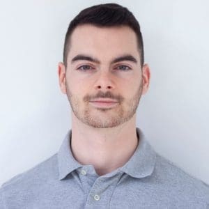
Dr Victor Racine
Founder & CEO of QuantaCell SAS
Hôpital Saint Eloi, IRMB, Montpellier
Thursday, April 30, 2026 at 11:00 AM
IECB auditorium, 2 rue Robert Escarpit, 33600 Pessac
Host: BioFAP Department of CBMN (M Raoux, A Bouter, O Lambert)
Summary:
Bio-imaging is rapidly expanding, from cell culture systems (2D/3D, organoids) to digital
histology, yet one major bottleneck remains: limited annotated data and time-consuming
analysis.
In this talk, Victor Racine will explore how foundation models are reshaping the field by
enabling powerful image analysis across scales:
Automated analysis in cell culture and high-content screening;
Robust feature extraction and tumor detection in histology;
Strong performance, even with limited data
Selected publications related to this presentation:
Ong et al. (2025) Digitalized organoids: integrated pipeline for high-speed 3D analysis of organoid
structures using multilevel segmentation and cellular topology. Nat Methods 22:1343–1354.
Domblides et al. (2024) Human NLRC4 expression promotes cancer survival and associates with type I
interferon signaling and immune infiltration. J Clin Invest 134:e166085.
Charrasse et al. (2023) Quantitative assessment of mitochondrial morphology relevant for studies on
cellular health and environmental toxicity. Comput Struct Biotechnol J. 21:5609-561.
Beghin et al. (2022) Automated high-speed 3D imaging of organoid cultures with multi-scale
phenotypic quantification. Nat Methods 19:881–892.
Blanc et al. (2020) Artificial intelligence solution to classify pulmonary nodules on CT. Diagn Interv
Imaging 101:803–810.
Frye et al. (2021) Mitochondrial morphology is associated with respiratory chain uncoupling in autism
spectrum disorder. Transl Psychiatry 11:527